| | You may also be interested in... |
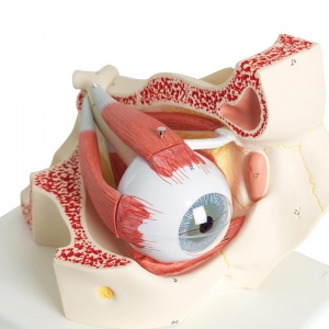
- Large anatomical model of the human eye in orbit
- Shows the optic nerve in its natural position (bony orbit of the eye)
- Can be disassembled into seven parts for further study
- Perfect for anatomical demonstrations in the classroom
 | | Usually delivered within 10 working days |
|
|
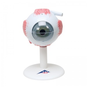
- Anatomical model of the eye in six parts, and three times life-size
- Dissects into both halves of sclera and choroid, lens and vitreous humour
- Larger size allows clear and educational demonstrations
- Several major structures can be studied in detail
| | Usually delivered within 10 working days |
|
|
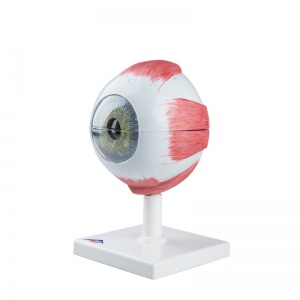
- Anatomical model of the eye in six parts, at five times life-size
- Sclera and choroid, lens and vitreous humour can all be removed
- Extra large size allows clear and educational demonstrations
- Several major structures can be studied in detail
| | Usually delivered within 10 working days |
|
|
|