Skin Model (3-Part)
The Skin Model is a useful model for teaching and learning about the anatomy and role of the skin. With an average surface area of over 21 square feet, skin is the largest organ of the body. This model helps to educate about the layers of the skin, and the important purposes they serve within the body.
About the SKin Model
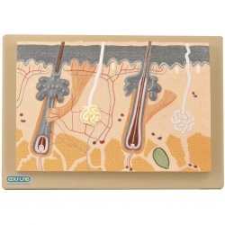
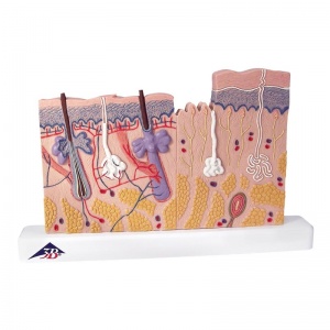
The skin is shown 80 times magnified, so even the smallest structures can be seen in detail. It can also be taken apart easily thanks to magnetic connectors, allowing for better demonstration. The Skin Model represents the three most important sections of skin: the human scalp, the palm of a hand, and the back of a hand.
All layers of the skin (epidermis, dermis and subcutis) and microscopic structures such as nerves, vessels and tactile corpuscles are shown in an anatomically accurate representation.
Contents of the Skin Model
The model is comprises of three parts, which are:
- Microanatomical representation of the papillary layer (e.g. localised in the palm of the hand)
- Microanatomical representation of the reticular layer (e.g. localised at the back of the hand)
- Longitudinal section through the human scalp with hair follicle and root sheath
Features of the Skin Model
- Three part anatomical model for learning about the human skin
- Helps to educate about the layers of the skin, and their roles
- All layers are shown in an anatomically accurate representation
- Skin is shown 80 times magnified, so even the smallest structures can be studied in detail
- Sections of human scalp, palm of a hand and the back of a hand
- Magnetic connections allow for the three parts to be taken apart easily and fast
Specifications of the Skin Model
- Dimensions: 34 x 39 x 15.5cm
- Weight: 2kg